|
Skupina vedená Oldřichem Zahradníčkem se zabývá biologickými účinky záření a radioterapií v následujích tématech:
- Radiační poškození DNA, proteinů a jejich komplexů
- Buněčná radiobiologie
- Biologické účinky nabitých částic, XUV a UV
- Teoretické modelování fyzikálního a chemického stádia interakce záření s biologickým materiálem ve vodě, vývoj stochastického kódu RADAMOL
- Charakterizace radioterapeutických svazků
- Fragmentace primárních iontů
Výběr z publikační činnosti
- Jelínek Michaelidesová, A., Kundrát, P., Zahradníček, O., Danilová, I., Pachnerová Brabcová, K., Vachelová, J., Vilimovský, J., David, M., Vondráček, V., Davídková, M. First independent validation of the proton-boron capture therapy concept. Scientific Reports. 14, (2024).
- Souli, M. et al. Clustered DNA damage patterns after proton therapy beam irradiation using plasmid DNA. Int. J. Mol. Sci. 23 (2022).
- Kraus, K. M. et al. Potential morbidity reduction for lung stereotactic body radiation therapy using respiratory gating. Cancers (Basel). 13, (2021).
- Simonetto, C. et al. Estimating long-term health risks after breast cancer radiotherapy: merging evidence from low and high doses. Radiat. Environ. Biophys. 60, 459–474 (2021).
- Kundrát, P., Friedland, W., Ottolenghi, A. & Baiocco, G. Coupling Radiation Transport and Track-Structure Simulations: Strategy Based on Analytical Formulas Representing DNA Damage Yields. Front. Phys. 9, 518 (2021).
- Tran, H. N. et al. Assessment of DNA damage with an adapted independent reaction time approach implemented in Geant4-DNA for the simulation of diffusion-controlled reactions between radio-induced reactive species and a chromatin fiber. Med. Phys. 48, 890–901 (2021).
- Wochnik, A. et al. Out-of-field doses for scanning proton radiotherapy of shallowly located paediatric tumours-a comparison of range shifter and 3D printed compensator. Phys. Med. Biol. 66, (2021).
- Port, M. et al. Gene Expression Changes in Irradiated Baboons: A Summary and Interpretation of a Decade of Findings. Radiation Research 195, 501–521 (2021).
- Sala, L. et al. Folding DNA into origami nanostructures enhances resistance to ionizing radiation. Nanoscale 13, 11197–11203 (2021).
- Krátká, M. et al. Gamma radiation effects on diamond field-effect biosensors with fibroblasts and extracellular matrix. Colloids Surfaces B Biointerfaces 204, 111689 (2021).
- Borm, K. J. et al. Toxicity of internal mammary irradiation in breast cancer. Are concerns still justified in times of modern treatment techniques? Acta Oncol. 59, 1201–1209 (2020).
- Kundrát, P. et al. Analytical formulas representing track-structure simulations on DNA damage induced by protons and light ions at radiotherapy-relevant energies. Sci. Reports 2020 101 10, 1–11 (2020).
- Hampl, M. et al. Polarized Sonic Hedgehog Protein Localization and a Shift in the Expression of Region-Specific Molecules Is Associated With the Secondary Palate Development in the Veiled Chameleon. Front. Cell Dev. Biol. 8, 572 (2020).
- Kavková, M. et al. Coordinated labio-lingual asymmetries in dental and bone development create a symmetrical acrodont dentition. Sci. Rep. 10, (2020).
- Torrisi, L., Davídková, M., Havránek, V., Cutroneo, M. & Torrisi, A. Physical study of proton therapy at CANAM laboratory on medulloblastoma cell lines DAOY. Radiat. Eff. Defects Solids 175, 863–878 (2020).
- Ostheim, P. et al. Overcoming challenges in human saliva gene expression measurements. Sci. Rep. 10, 1–12 (2020).
- Henthorn, N. T. et al. Mapping the Future of Particle Radiobiology in Europe: The INSPIRE Project. Front. Phys. 8, 438 (2020).
- Cruz-Garcia, L. et al. In vivo validation of alternative FDXR transcripts in human blood in response to ionizing radiation. Int. J. Mol. Sci. 21, 1–18 (2020).
- Michaelidesová, A. et al. In vitro comparison of passive and active clinical proton beams. Int. J. Mol. Sci. 21, 1–15 (2020).
- Kundrát, P. et al. What anatomic features govern personal long-term health risks from breast cancer radiotherapy? Radiat. Prot. Dosimetry 186, 381–385 (2019).
- Friedland, W., Kundrát, P., Becker, J. & Eidemüller, M. Biophysical simulation tool PARTRAC: Modelling proton beams at therapy-relevant energies. Radiat. Prot. Dosimetry 186, 172–175 (2019).
- Romanenko, O. et al. Performance and application of heavy ion nuclear microbeam facility at the Nuclear Physics Institute in Řež, Czech Republic. Rev. Sci. Instrum. 90, 013701 (2019).
- Schuemann, J. et al. A New Standard DNA Damage (SDD) Data Format. Radiat. Res. 191, 76–92 (2019).
- Pachnerová Brabcová, K., Sihver, L., Ukraintsev, E., Štěpán, V. & Davídková, M. How detection of plasmid DNA fragmentation affects radiation strand break yields. Radiat. Prot. Dosimetry 183, 89–92 (2019).
- Ondrák, L. et al. Radioprotective effect of hydroxyl radical scavengers on prokaryotic and eukaryotic cells under various gamma irradiation conditions. Radiat. Prot. Dosimetry 186, 186–190 (2019).
- Pachnerová Brabcová, K. et al. Radiation-induced plasmid DNA damage: Effect of concentration and length. Radiat. Prot. Dosimetry 186, 168–171 (2019).
- Konířová, J. et al. Differentiation Induction as a Response to Irradiation in Neural Stem Cells In Vitro. Cancers (Basel). 11, 913 (2019).
- Michaelidesová, A. et al. Comparison of the radiation sensitivity of the breast cancer cell line MCF7 and adipose-derived stem cells. Radiat. Prot. Dosimetry 186, 155–158 (2019).
- Michaelidesová, A., Konířová, J., Bartůněk, P. & Zíková, M. Effects of radiation therapy on neural stem cells. Genes vol. 10 640 (2019).
- Tichy, A. et al. The first in vivo multiparametric comparison of different radiation exposure biomarkers in human blood. PLoS One 13, (2018).
- Vyšín, L. et al. Biological action in and out of the water window. Acta Phys. Pol. A 133, 236–238 (2018).
- Stolarczyk, L. et al. Dose distribution of secondary radiation in a water phantom for a proton pencil beam - EURADOS WG9 intercomparison exercise. Phys. Med. Biol. 63, (2018).
- Ježková, L. et al. Particles with similar LET values generate DNA breaks of different complexity and reparability: A high-resolution microscopy analysis of γH2AX/53BP1 foci. Nanoscale 10, 1162–1179 (2018).
- Pachnerová Brabcová, K., Sihver, L., Ukraintsev, E., Štěpán, V. & Davídková, M. Length computation of irradiated plasmid DNA molecules. Biointerphases 13, 061005 (2018).
- Vyšín, L. et al. Dose-Rate Effects in Breaking DNA Strands by Short Pulses of Extreme Ultraviolet Radiation. Radiat. Res. 189, 466–476 (2018).
- Šefl, M., Pachnerová Brabcová, K. & Štěpán, V. Dosimetry as a Catch in Radiobiology Experiments. Radiat. Res. 190, 404–411 (2018).
- Reimitz, D., Davídková, M., Mestek, O., Pinkas, J. & Kočišek, J. Radiomodifying effects of RAPTA C and CDDP on DNA strand break induction. Radiat. Phys. Chem. 141, 229–234 (2017).
- Vyšín, L. et al. Degradation of phospholipids under different types of irradiation and varying oxygen saturation. Radiat. Environ. Biophys. 56, 241–247 (2017).
- Michaelidesová, A. et al. Relative biological effectiveness in a proton spread-out Bragg peak formed by pencil beam scanning mode. Australas. Phys. Eng. Sci. Med. 40, 359–368 (2017).
- Štěpán, V. & Davídková, M. Understanding radiation damage on sub-cellular scale using RADAMOL simulation tool. Radiat. Phys. Chem. 128, 11–17 (2016).
- Adjei, D. et al. DNA strand breaks induced by soft X-ray pulses from a compact laser plasma source. Radiat. Phys. Chem. 120, 17–25 (2016).
- Krátká, M. et al. Gamma radiation effects on hydrogen-terminated nanocrystalline diamond bio-transistors. Diam. Relat. Mater. 63, 186–191 (2016).
- Stursa, J. et al. Mass production of fluorescent nanodiamonds with a narrow emission intensity distribution. Carbon N. Y. 96, 812–818 (2016).
- Marshall, T. I. et al. Investigating the Implications of a Variable RBE on Proton Dose Fractionation Across a Clinical Pencil Beam Scanned Spread-Out Bragg Peak. Int. J. Radiat. Oncol. Biol. Phys. 95, 70–77 (2016).
- Pachnerová Brabcová, K. et al. Contribution of indirect effects to clustered damage in DNA irradiated with protons. Radiat. Prot. Dosimetry 166, 44–48 (2015).
- Vyšín, L. et al. Proton-induced direct and indirect damage of plasmid DNA. Radiat. Environ. Biophys. 54, 343–352 (2015).
- Bernal, M. A. et al. A review of the Geant4-DNA very low energy extension of the Geant4 Monte Carlo simulation toolkit. Phys. Medica 31, 861–874 (2015).
- Nováková, E. et al. Breaking DNA strands by extreme-ultraviolet laser pulses in vacuum. Phys. Rev. E - Stat. Nonlinear, Soft Matter Phys. 91, 042718 (2015).
- Litvinchuk, A. V., Vachelová, J., Michaelidesová, A., Wagner, R. & Davídková, M. Dose-dependent micronuclei formation in normal human fibroblasts exposed to proton radiation. Radiat. Environ. Biophys. 54, 327–34 (2015).
- Falk, M. et al. Determining omics spatiotemporal dimensions using exciting new nanoscopy techniques to assess complex cell responses to DNA damage: Part B-structuromics. Crit. Rev. Eukaryot. Gene Expr. 24, 225–247 (2014).
- Falk, M. et al. Primary and secondary clustering of DSB repair foci and repair kinetics compared for X-rays, protons of different energies and high-LET 20Ne ions. J. Radiat. Res. 55, i79–i80 (2014).
- Karamitros, M. et al. Diffusion-controlled reactions modeling in Geant4-DNA. J. Comput. Phys. 274, 841–882 (2014).
- Ježková, L. et al. Function of chromatin structure and dynamics in DNA damage, repair and misrepair: γ-rays and protons in action. Appl. Radiat. Isot. 83, 128–136 (2014).
- Incerti, S. et al. Simulating radial dose of ion tracks in liquid water simulated with Geant4-DNA: A comparative study. Nucl. Instruments Methods Phys. Res. Sect. B Beam Interact. with Mater. Atoms 333, 92–98 (2014).
- Falk, M. et al. Chromatin differentiation of white blood cells decreases DSB damage induction, prevents functional assembly of repair foci, but has no influence on protrusion of heterochromatic DSBs into the low-dense chromatin. J. Radiat. Res. 55, i81–i82 (2014).
- Pachnerová Brabcová, K. et al. Clustered DNA damage on subcellular level: effect of scavengers. Radiat. Environ. Biophys. 53, 705–712 (2014).
- Štěpán, V. & Davídková, M. RADAMOL tool: Role of radiation quality and charge transfer in damage distribution along DNA oligomer Guest editors: Andrey V. Solov’yov, Nigel Mason, Paulo Limão-Vieira, Malgorzata Smialek-Telega. Eur. Phys. J. D 68, 1–7 (2014).
- Adjei, D. et al. Development of a compact laser-produced plasma soft X-ray source for radiobiology experiments. Nucl. Instruments Methods Phys. Res. Sect. B Beam Interact. with Mater. Atoms 364, 27–32 (2014).
- Jezkova, L. et al. Function of chromatin structure and dynamics in DNA damage, repair and misrepair: gamma-rays and protons in action. Appl. Radiat. Isot. 83 Pt B, 128–136 (2014).
- Falk, M. et al. Heterochromatinization associated with cell differentiation as a model to study DNA double strand break induction and repair in the context of higher-order chromatin structure. Appl. Radiat. Isot. 83, 177–185 (2014).
- Falk, M. et al. Determining Omics spatiotemporal dimensions using exciting new nanoscopy techniques to assess complex cell responses to DNA damage: part A--radiomics. Crit Rev Eukaryot Gene Expr 24, 205–223 (2014).
- Staaf, E. et al. Characterisation of a setup for mixed beam exposures of cells to 241Am alpha particles and X-rays. Radiat. Prot. Dosimetry 151, 570–579 (2012).
- Staaf, E. et al. Micronuclei in human peripheral blood lymphocytes exposed to mixed beams of X-rays and alpha particles. Radiat. Environ. Biophys. 51, 283–293 (2012).
- Nováková, E. et al. Damage to dry plasmid DNA induced by nanosecond XUV-laser pulses. in Damage to VUV, EUV, and X-ray Optics III vol. 8077 80770W (SPIE, 2011).
- Spotheim-Maurizot, M. & Davídková, M. Radiation damage to DNA in DNA-protein complexes. Mutat. Res. - Fundam. Mol. Mech. Mutagen. 711, 41–48 (2011).
- Francis, Z. et al. Molecular scale track structure simulations in liquid water using the Geant4-DNA Monte-Carlo processes. Appl. Radiat. Isot. 69, 220–226 (2011).
|
|
 Jana pracuje v laminárním boxu. Jana pracuje v laminárním boxu.
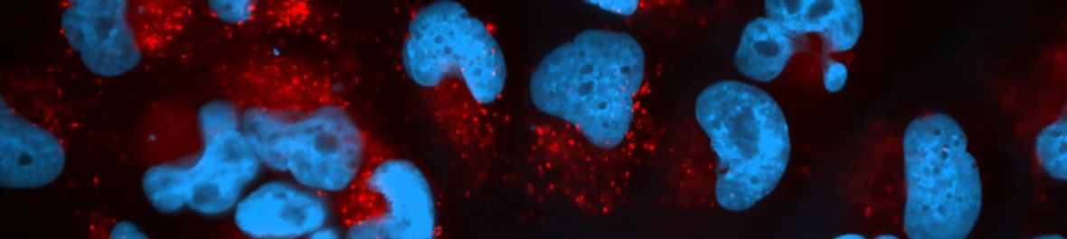
 Anna si ohromeně prohlíží ozářené fibroblasty mikroskopem. Anna si ohromeně prohlíží ozářené fibroblasty mikroskopem.

Richard kontroluje ionizační komoru před ozářením biologických vzorků.

Pohled na laboratorní stoly zářící novotou.

Agarózové gely se vzorky DNA na UV stolku.
|